Home » Without Label » Tendon Diagram / 3 Schematic representation of the hierarchical ... / Fpe medical review board a foot pain diagram is a great tool to help you work out what is causing your ankle and foot pain.
Tendon Diagram / 3 Schematic representation of the hierarchical ... / Fpe medical review board a foot pain diagram is a great tool to help you work out what is causing your ankle and foot pain.
Tendon Diagram / 3 Schematic representation of the hierarchical ... / Fpe medical review board a foot pain diagram is a great tool to help you work out what is causing your ankle and foot pain.. Again, our knowledge of how mechanical stimulus mediates ligament and tendon structure is more empirical and less. Ligaments join the knee bones and provide stability to the knee: The fcu tendon is one of two tendons that bend the wrist. Diagram of inside the body. Possibly the most important tendon in terms of mobility is the achilles tendon.
Tendons are the connection between bones and muscles tendon diagram. 9 photos of the foot tendons and ligaments diagram. Tendons, located at each end of a muscle, attach muscle to bone. The largest of these shoulder muscles is the. Posted on april 3, 2019april 3, 2019.
Osgood-Schlatter Disease | Johns Hopkins Medicine Health ... from www.hopkinsmedicine.org The anterior cruciate ligament prevents the femur from sliding backward on the tibia (or the tibia sliding forward on the femur). Possibly the most important tendon in terms of mobility is the achilles tendon. The lower part of the trapezius ascends and depresses the scapula, while the transverse or middle region of the trapezius is what retracts the. This muscle diagram is interactive: If you tear the biceps tendon at the shoulder, you may lose some strength in your arm and have pain when you forcefully turn your arm from palm down to palm up. Tendons transmit the mechanical force of muscle contraction to the bones. Ligaments and tendons are fibrous connective tissues made up of densely packed collagen fibers. The two peroneal tendons in the foot run side by side behind the outer ankle bone.
Ligaments and tendons are adapted in response to changes in mechanical stiffness.
By connecting our rigid bones to our powerful muscles, tendons allow us to move. Ligaments and tendons are adapted in response to changes in mechanical stiffness. The ecu tendon works along with the ecrl and ecrb to straighten the wrist. The pubis, ischium, and ilium together constitute the pelvis while the thigh bone is the femur. Diagram of tendons in forearm. Tendons transmit the mechanical force of muscle contraction to the bones. The achilles tendon is a tough band of fibrous tissue that connects the calf muscles to the heel bone (calcaneus). If the tendon cannot be identified then a complete tear of the tendon should be sought. Movement occurs when our muscles pull on our bones, relocating them. The bones together make up the hip. Also allows the action of raising up onto toes. Allows the action of raising the foot. Possibly the most important tendon in terms of mobility is the achilles tendon.
Ligaments and tendons are adapted in response to changes in mechanical stiffness. If the tendon cannot be identified then a complete tear of the tendon should be sought. The fcu tendon is one of two tendons that bend the wrist. Movement occurs when our muscles pull on our bones, relocating them. Your biceps tendons attach the biceps muscle to bones in the shoulder and in the elbow.
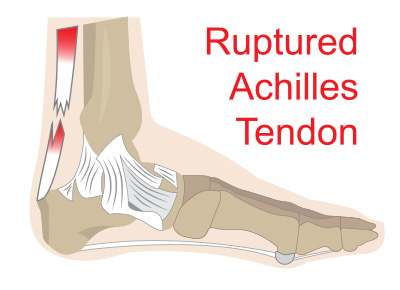
Achilles Tendon Ruptures | Issaquah Foot & Ankle Specialists from images.fosterwebmarketing.com Your biceps tendons attach the biceps muscle to bones in the shoulder and in the elbow. Foot anatomy diagram, foot joint diagram, foot sprain diagram, foot tendons and ligaments pain, leg tendon diagram, peroneal tendonitis, foot, foot anatomy diagram, foot joint diagram, foot sprain diagram, foot tendons and ligaments pain, leg tendon diagram, peroneal tendonitis. The bones of the hip include the femur, the ilium, the ischium, and the pubis. The coracobrachialis muscle lies deep to the biceps brachii in the arm. The achilles tendon enables us to walk, without it we would not be able to raise our heels of the ground. Tendons, located at each end of a muscle, attach muscle to bone. Tendons are similar to ligaments; The anterior cruciate ligament prevents the femur from sliding backward on the tibia (or the tibia sliding forward on the femur).
Tendon diagram of the knee.
Tendons are the connection between bones and muscles. Diagram of tendons in forearm. Foot anatomy diagram, foot joint diagram, foot sprain diagram, foot tendons and ligaments pain, leg tendon diagram, peroneal tendonitis, foot, foot anatomy diagram, foot joint diagram, foot sprain diagram, foot tendons and ligaments pain, leg tendon diagram, peroneal tendonitis. Tendon diagram simple / 8.4c: If you tear the biceps tendon at the shoulder, you may lose some strength in your arm and have pain when you forcefully turn your arm from palm down to palm up. The bones together make up the hip. Tendons, located at each end of a muscle, attach muscle to bone. 9 photos of the foot tendons and ligaments diagram. Attaches the calf muscles to the calcaneus, most important muscles for running, jumping, walking etc. Ligaments and tendons are fibrous connective tissues made up of densely packed collagen fibers. Tendon, tissue that attaches a muscle to other body parts, usually bones. The changes in ligaments and tendons generally occur more slowly than adaptation in bone, because ligaments and tendons have less vascular supply. Both are made of collagen.ligaments connect one bone to another, while tendons connect muscle to bone.
You can see a diagram of the achilles tendon below. Tendons are found throughout the body, from the head and neck all the way down to the feet. Tendons, located at each end of a muscle, attach muscle to bone. The trapezius or trapezoid muscles are two paired muscles that extend from the base of the thoracic vertebrae in the spine to the occipital bone and run out to the spine of the scapula. This important tendon in the back of the calf and ankle connects the plantaris, gastrocnemius, and soleus muscles to.
begin to dig: Mick Wilkinson Part I: Why Barefoot Running ... from 1.bp.blogspot.com It attaches to the wrist bone, the pisiform, and as well as the 5th hand bone. The two peroneal tendons in the foot run side by side behind the outer ankle bone. Again, our knowledge of how mechanical stimulus mediates ligament and tendon structure is more empirical and less. Tendons are the connection between bones and muscles tendon diagram. The changes in ligaments and tendons generally occur more slowly than adaptation in bone, because ligaments and tendons have less vascular supply. The pubis, ischium, and ilium together constitute the pelvis while the thigh bone is the femur. The bones of the hip include the femur, the ilium, the ischium, and the pubis. There are three parts to the trapezius.
Learn about these muscles, their origin and insertion points, and their functional anatomy.
Tendons, located at each end of a muscle, attach muscle to bone. Tendons attach muscles to bones. The ecu tendon works along with the ecrl and ecrb to straighten the wrist. Anatomy of leg muscles and tendons anatomy diagram leg muscles and tendons anatomy diagram pics photo, anatomy of leg muscles and tendons anatomy diagram leg muscles. Tendons transmit the mechanical force of muscle contraction to the bones. Attaches the calf muscles to the calcaneus, most important muscles for running, jumping, walking etc. When the muscles tighten (contract) arguably, the most important tendon is the achilles tendon, which allows the calf muscles to move. This results in collapse of the arch of the foot (commonly called flatfoot or flat foot), along with foot and sometimes ankle deformities that can become debilitating or disabling in later stages. The achilles tendon is the strongest and largest tendon in the body. The fcu tendon is one of two tendons that bend the wrist. Ligaments and tendons are adapted in response to changes in mechanical stiffness. On the other hand, the insertion is where a tendon attaches that muscle to the *more* movable bone. The two peroneal tendons in the foot run side by side behind the outer ankle bone.